
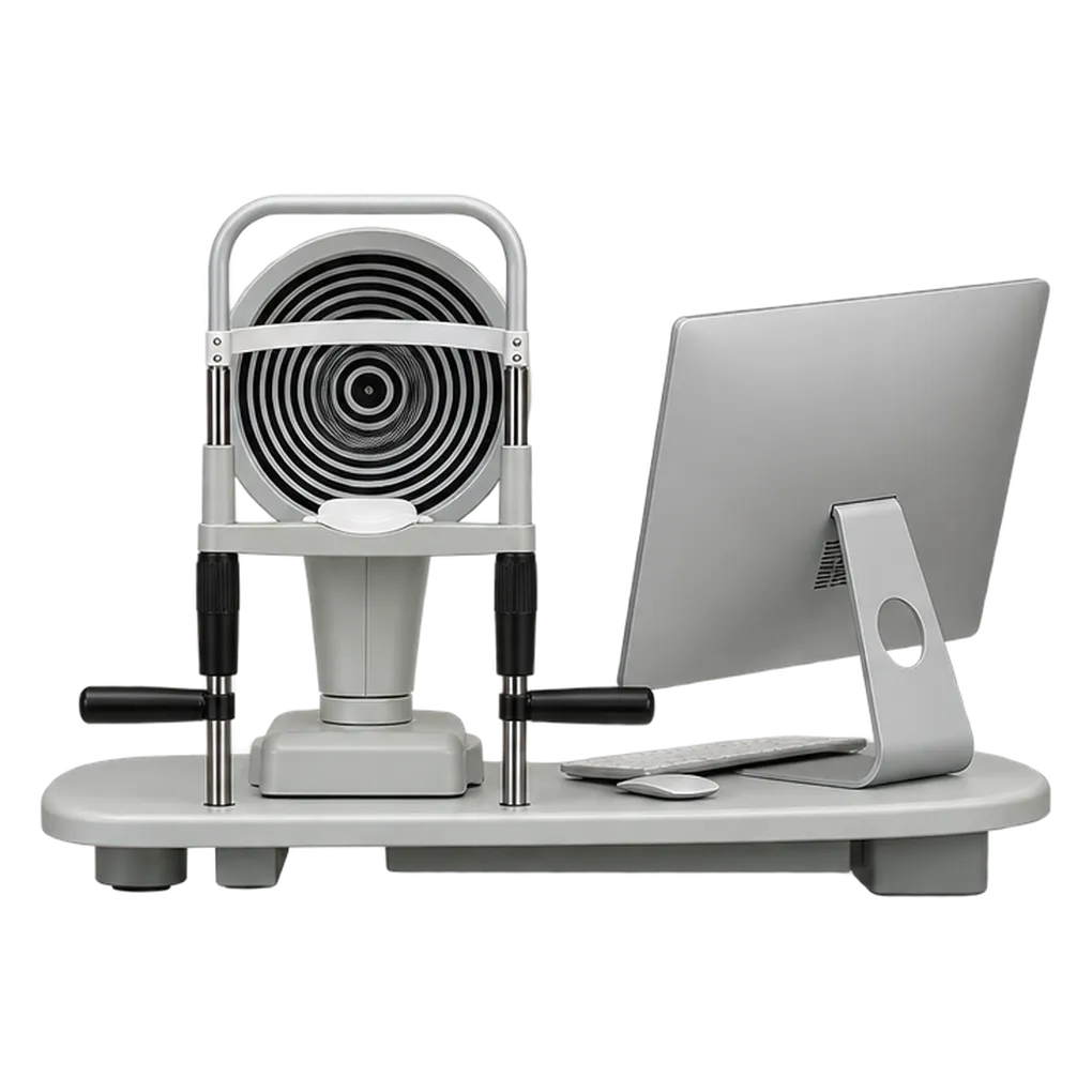
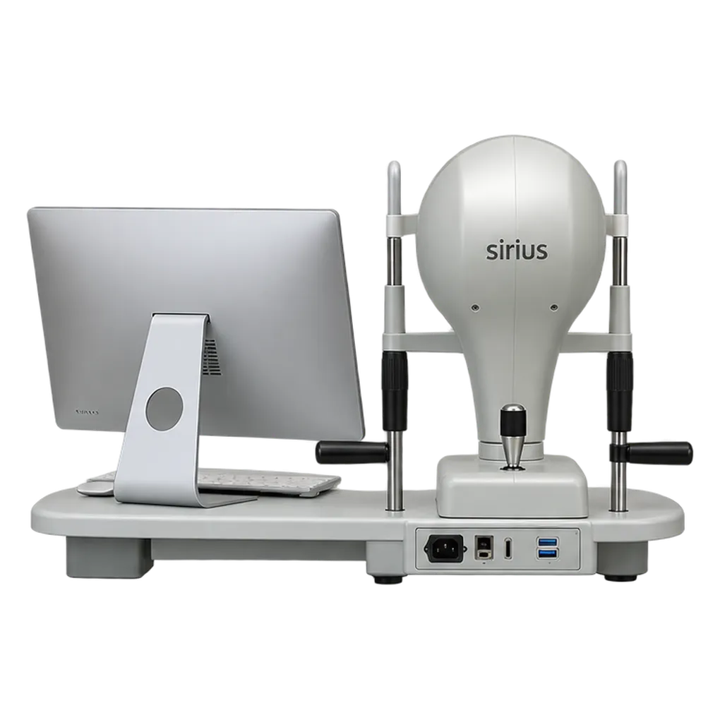
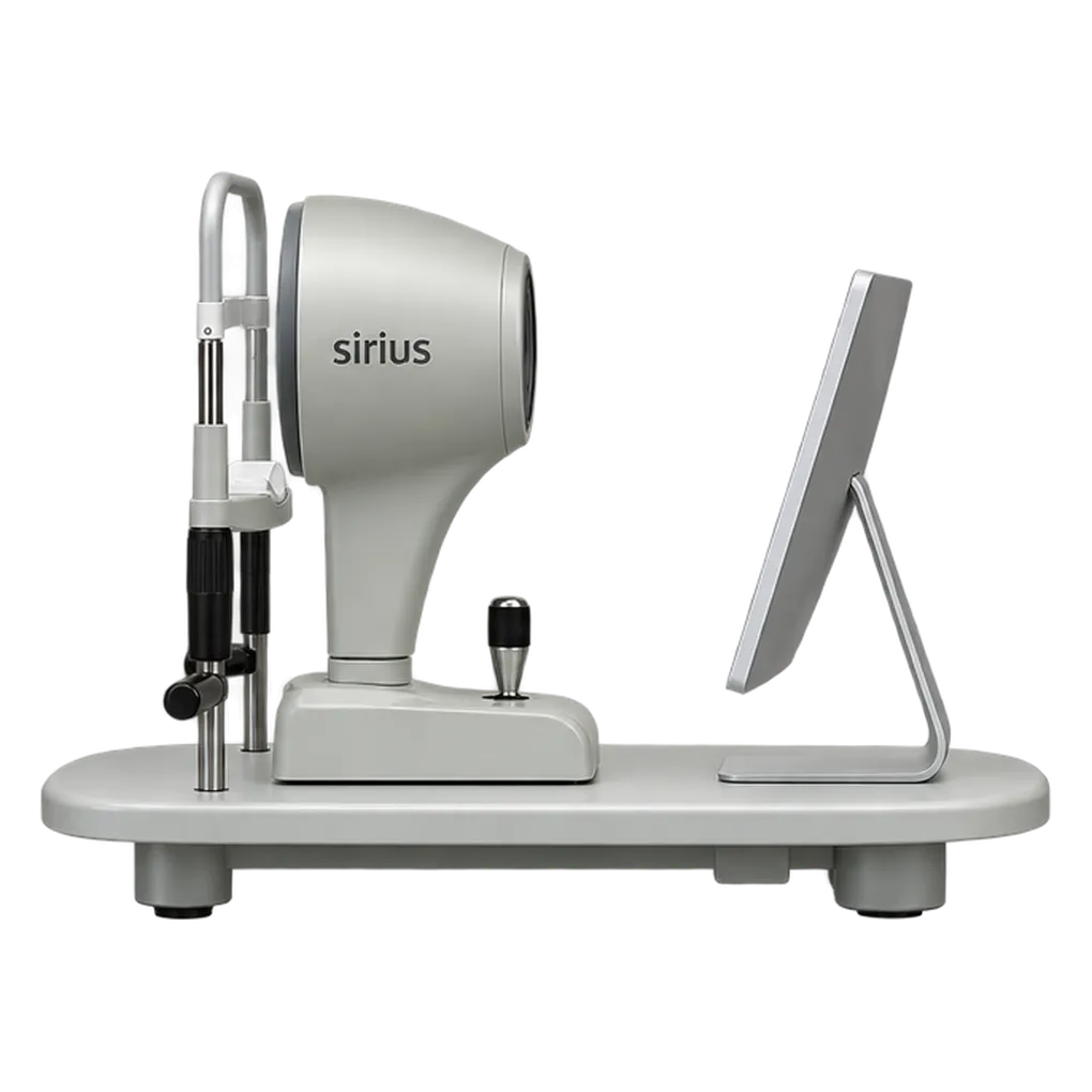
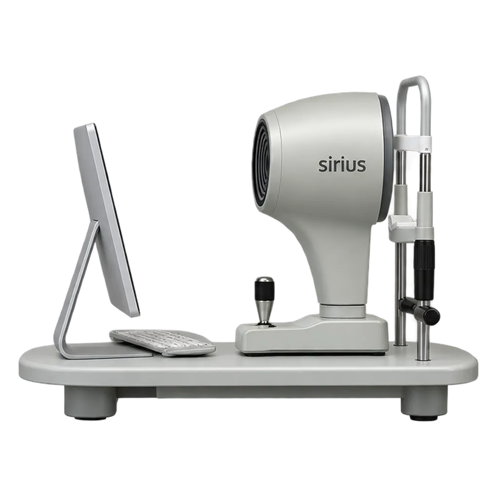
Refractive surgery practices, cataract clinics, glaucoma services, contact lens practices and dry eye clinics use the Sirius to examine the anterior segment. Ophthalmologists, optometrists and ophthalmic technicians operate it, and the results inform screening before corneal surgery, lens selection and ocular surface assessment.
The Sirius pairs Placido topography with Scheimpflug tomography, measuring the front and back corneal surfaces together. Pachymetry, elevation, curvature and dioptric power maps cover 12 mm, and high-resolution corneal sections feed anterior chamber biometry. A practice screening a candidate for refractive surgery reads thickness, elevation and keratoconus screening from the same examination.
Cataract work uses the optional ray-tracing module to calculate spherical and toric intraocular lens power from the measured cornea. Glaucoma services use the iridocorneal angle with pachymetry for screening. Corneal aberrometry at different pupil diameters, with OPD, point spread and modulation transfer simulations, describes how the cornea shapes the retinal image.
Contact lens practices simulate rigid lens fits from an internal database and use videokeratoscopy with 470 nm fluorescein lighting for rigid and orthokeratology lenses. Dry eye clinics combine non-invasive tear break-up time, infrared meibography and the dry eye report. Pupillography in scotopic, mesopic and photopic light adds a dynamic record.
Calibration is checked every 30 days with the supplied 8 mm radius sphere, and cleaning follows a set routine: parts away from the patient daily, and patient-contact parts after each use. Phoenix software stores each examination and shares it across CSO devices, and DICOM v3 compatibility under the IHE Eye Care profile lets results join an imaging workflow. At 7 kg, the head sits on a consulting-room table or an optional ophthalmic table.