

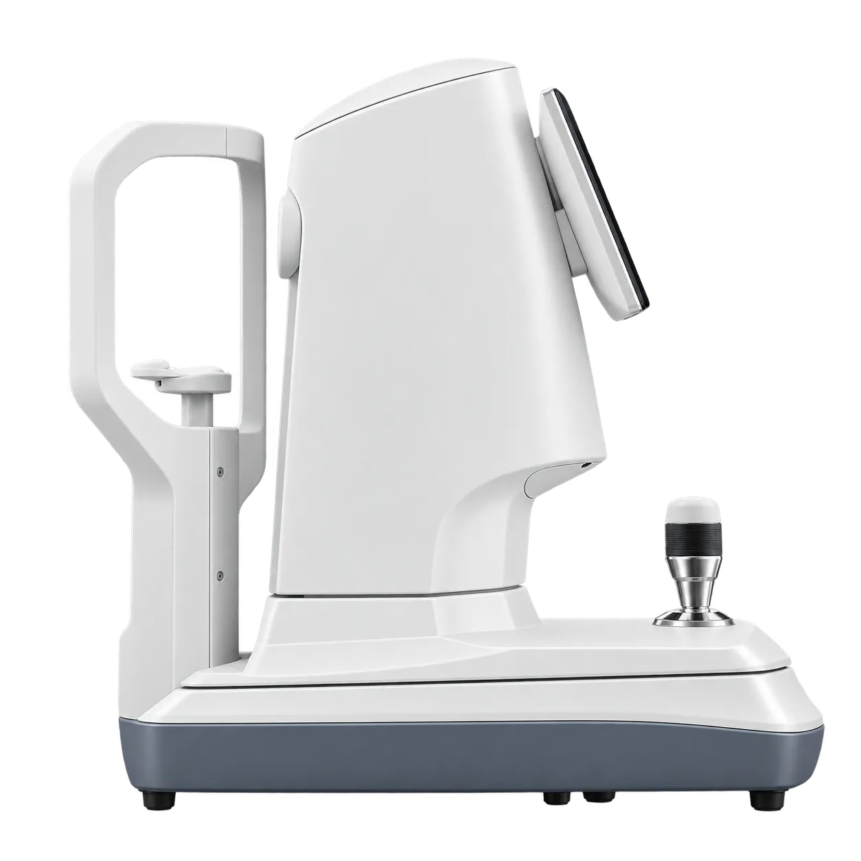
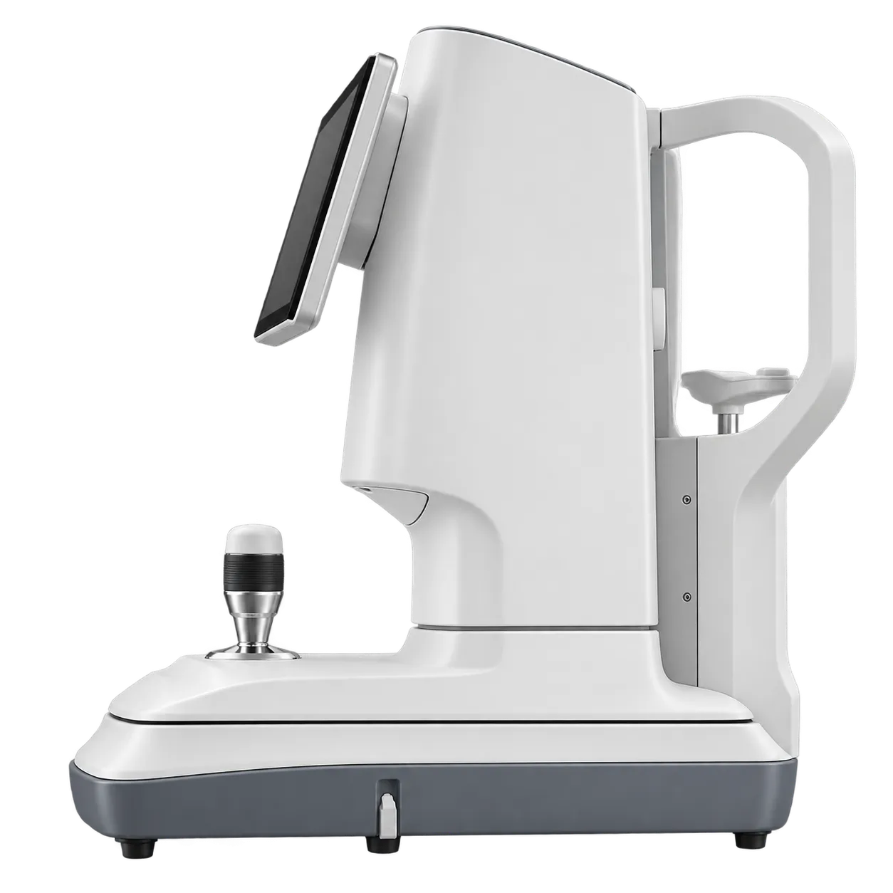
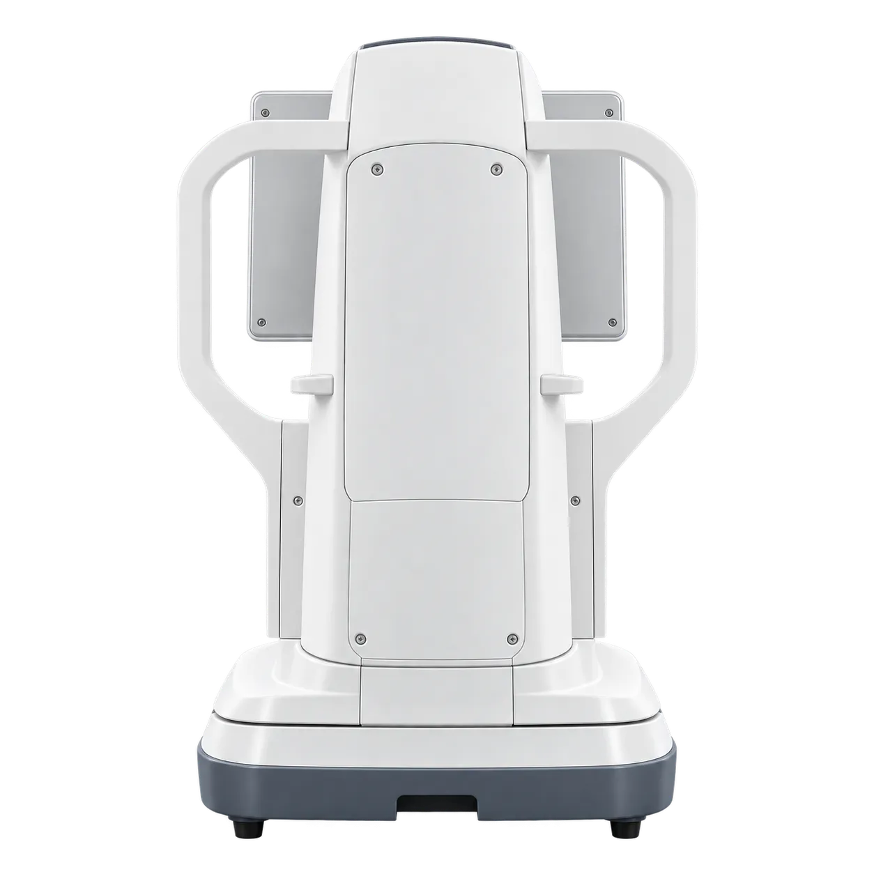
Dry eye clinics, contact lens practices, cornea services and general ophthalmology and optometry practices use the DEA520 to examine the ocular surface. Optometrists, ophthalmologists, contact lens practitioners and trained technicians work through the same screen, so a patient moves from the questionnaires to the imaging steps without changing instruments. A busy ocular surface clinic can plan a visit around one station, with the chin rest, screen, and report all within the examiner's reach.
For dry eye assessment, the built-in OSDI, McMonnies, SPEED and DEQ-5 questionnaires start the record. The lipid layer video with MGD templates, the eyelid margin zoom, the fluorescein staining view, the infrared blink analysis and the AI-assisted tear film break-up time follow in turn. Blink frequency, interval, incomplete blinks and their ratio are all reported, which gives a clinician measured values to discuss with the patient. Each result is stored in the same record as the imaging so that the clinician can return to it at the next appointment.
Corneal topography serves refractive, contact lens and orthokeratology work. The four maps, K1, K2, Km, the shape factors and sagittal height describe the front surface of the cornea, and Zernike wavefront aberration adds a PSF map, an MTF curve and simulated vision. RGP fitting with fluorescein simulation and orthokeratology recommendations then support the lens choice, and the orthokeratology function was developed with team SOS from the Fudan University Eye & ENT Hospital.
At the end of the visit, the report can be printed, exported to PDF or sent to a DICOM server. Patient and case management with database backup keeps follow-up examinations together, and case comparison with difference maps shows how a cornea has changed between visits.