
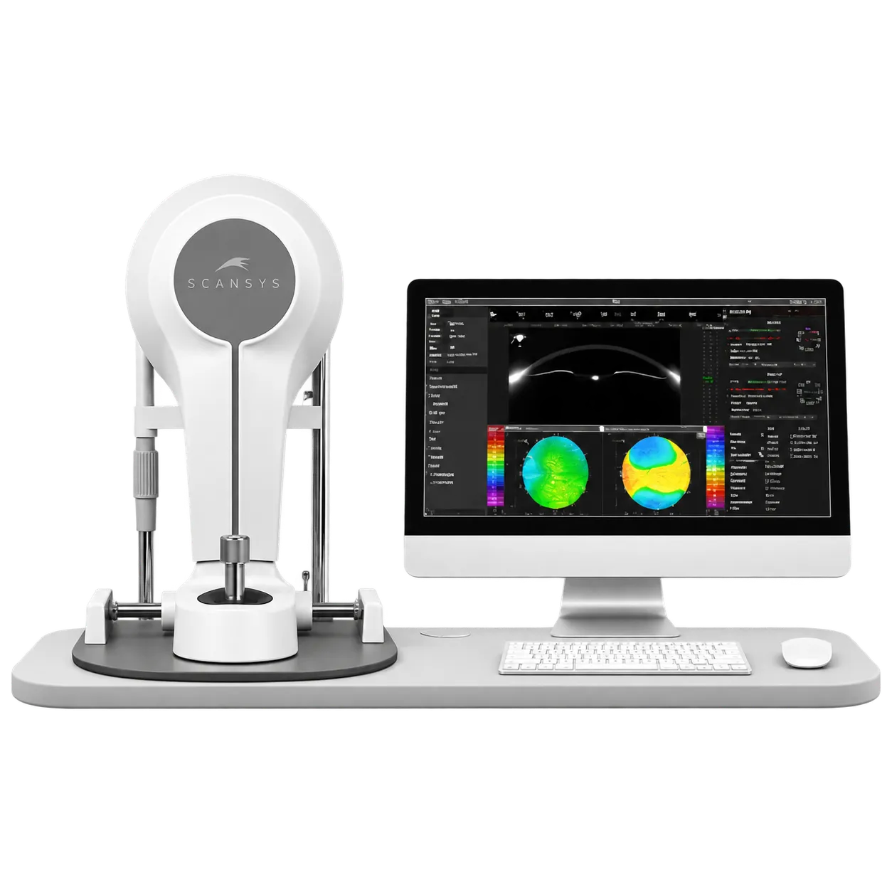
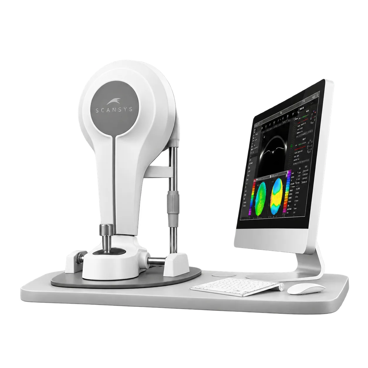
Refractive surgery practices, cataract clinics, glaucoma services and contact lens practices use the SCANSYS TA517 to examine the anterior segment. Ophthalmologists, optometrists and ophthalmic technicians run the scan, and the results inform screening before surgery, lens selection and fitting.
The capture is quick, which suits a busy list. A scan takes about one second and gathers 28 or 60 images, so each patient holds still only briefly, and the software builds a 3D model with elevation, thickness and refractive maps from that one capture. Binocular comparison of the Refractive 4 Maps and a two-examination comparison help at follow-up.
Refractive practices read keratoconus index, probability and grade from 0 to IV, with corneal thickness profile and shape factors beside them. Cataract clinics use corneal power values, total corneal astigmatism and spherical aberration, with the optional IOL module running Jin, SRK/T, Holladay 1 and Hoffer Q formulas. ICL planning adds a size recommendation and a vault prediction.
Glaucoma services use AOD, TISA, chamber angle and anterior chamber parameters for screening, with corneal thickness supporting IOP correction formulas. Contact lens practices fit orthokeratology, soft and RGP lenses from simulated fluorescein images, and the optional scleral lens module scans a 16 mm corneoscleral area.
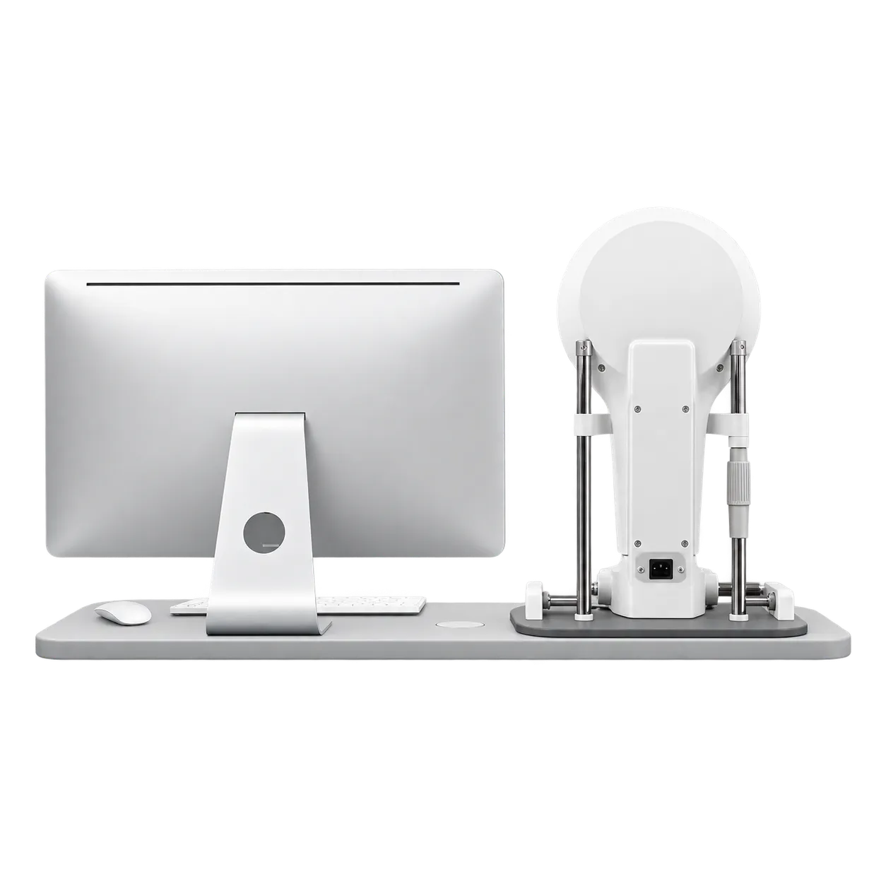
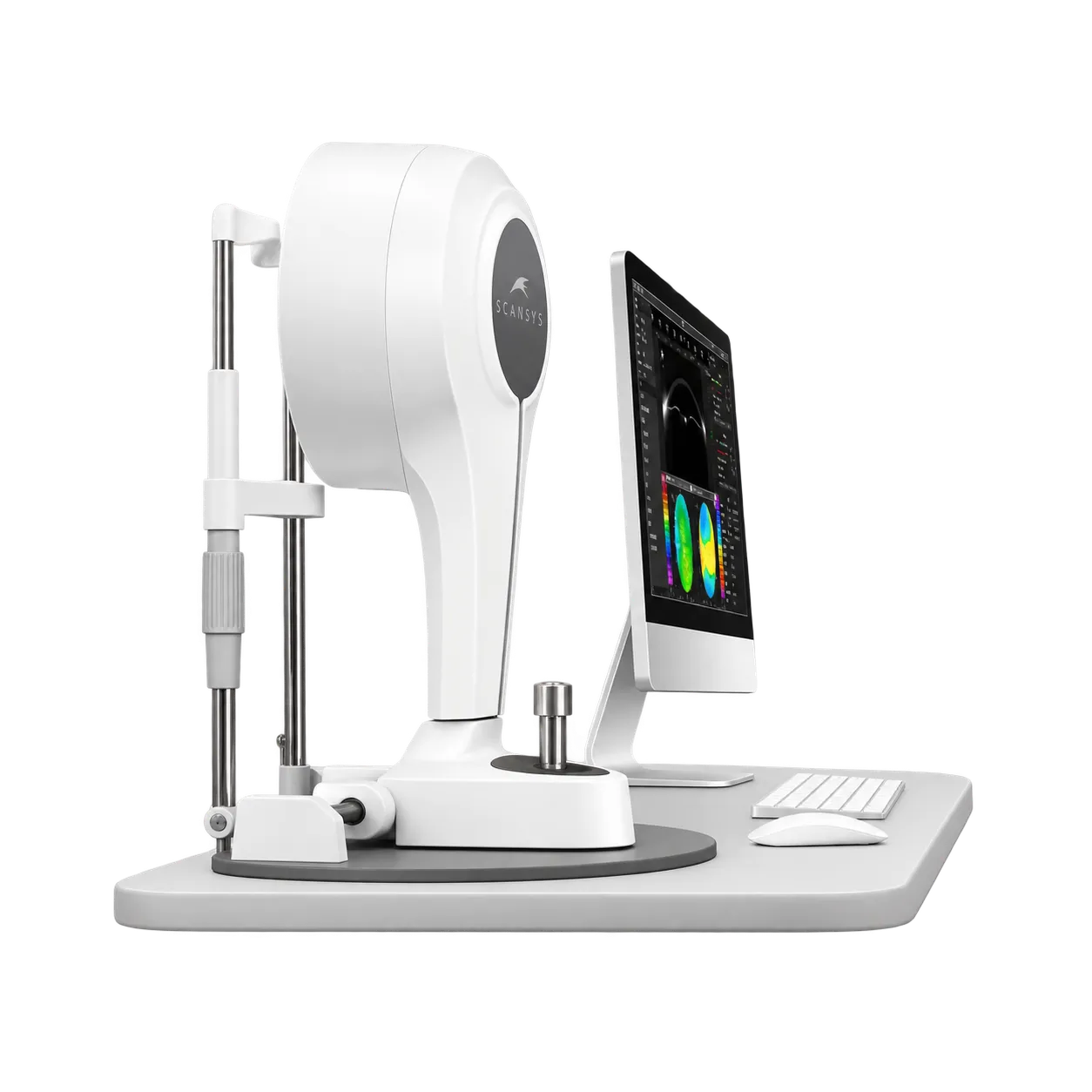
At 12 kg, the instrument sits on a stable table or an optional instrument table in a consulting or pre-test room. The working range gives 115 mm of travel front to back, 100 mm side to side and 30 mm up and down, which lets the operator centre each eye without moving the patient far. Results are reviewed on the connected computer, which runs the Scansys software on Windows 10, and two examinations can be shown together so that a change between visits is visible at the consultation.