
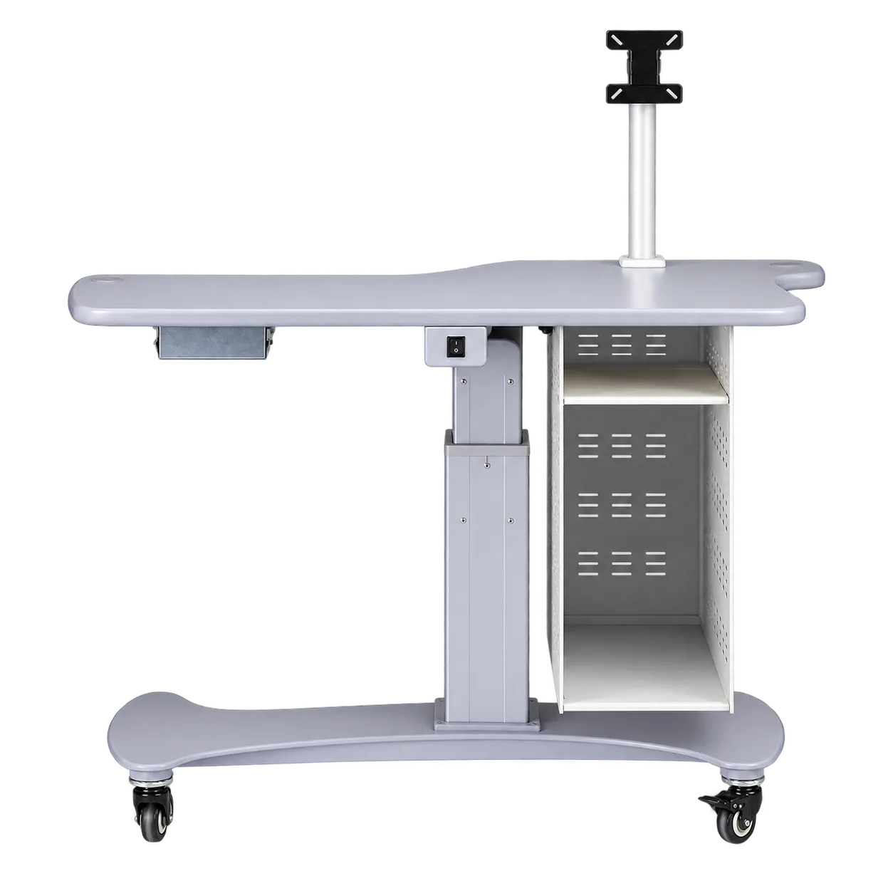
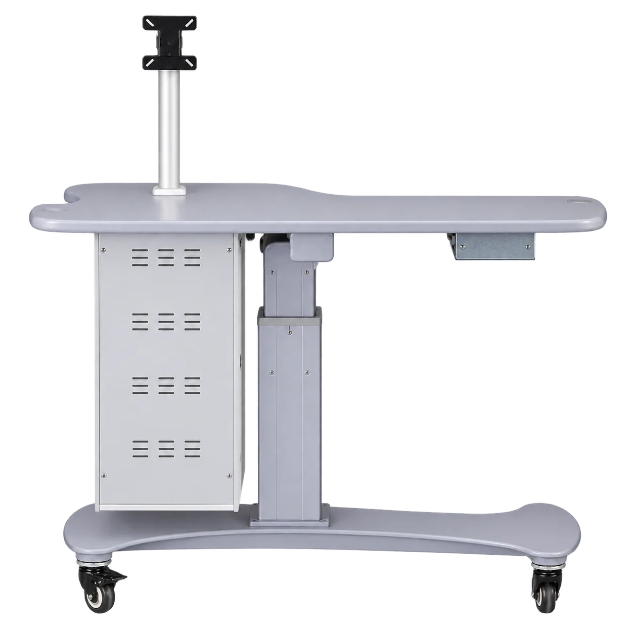
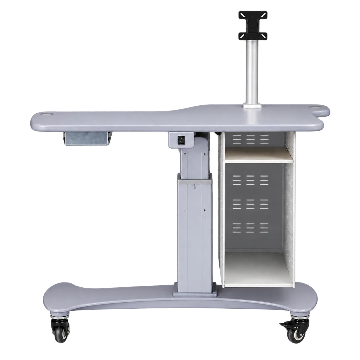
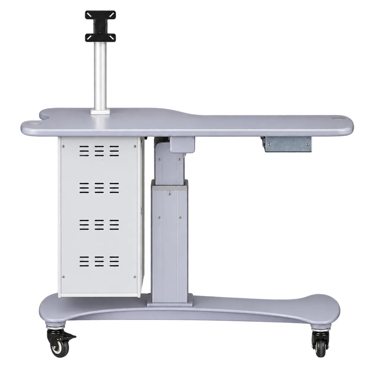
Imaging rooms in community practices are the natural home. OCT and retinal photography are now widely offered by UK optometrists, and each system brings a computer, a screen and cabling that otherwise spill across a desk. The C-280 gathers them into one station, keeping the room tidy and the cables off the floor.
Multi-room practices sharing one scanner benefit from the castors. A single OCT or camera can be wheeled to wherever it is needed that day, instead of patients being walked to a dedicated room. Secure cables and apply the brakes, once confirmed, before each use.
Hospital eye service imaging suites and medical retina and glaucoma clinics can use it to standardise imaging positions, with the same layout at every station so photographers and technicians find everything in the same place.
Retinal screening sessions held in shared or temporary rooms suit a mobile station. The table is furniture, not a screening device, so the camera and protocols remain those your programme approves.
Enhanced community services, such as minor eye conditions, glaucoma referral refinement and diabetic monitoring, increasingly rely on imaging to document findings. A dedicated imaging station supports the thorough record-keeping the GOC expects.
Corneal topography and contact lens fitting can use the station for a topographer and its computer. Digital slit lamps with attached cameras also benefit from the display mounted at the operator's eye line.
University optometry clinics can use it to give students a complete, self-contained imaging position.
Wheelchair users are partly served. Many imaging devices can be used from a wheelchair, but the C-280's column, cabinet, base and short height range limit clearance, so practices with many wheelchair users usually keep an accessible imaging position as well.