
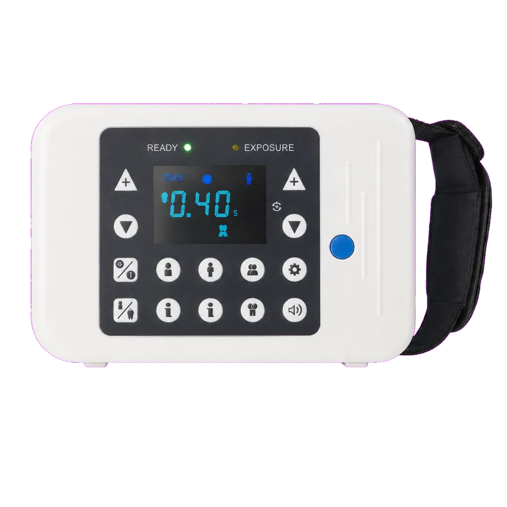
Intraoral radiography carries the diagnostic load in UK general dentistry, and the SJD-112 is configured for that daily volume. It supports bitewing caries detection, apical assessment of pathology across anterior, premolar and molar regions, endodontic working-length and obturation verification, periodontal bone-level review, and root morphology assessment ahead of extraction.
Multi-operator settings are the clearest fit. NHS practices running several surgeries, corporate groups, teaching clinics and community dental services all share equipment across clinicians, associates, therapists and nurses. Where technique varies between operators, image quality audits become hard to interpret and repeat rates drift upward. A control face organised by anatomical region gives every operator the same starting point.
The exposure envelope supports that structure. Molar regions sit at the higher end because of tissue thickness; apical views on slender anterior roots sit lower; paediatric patients lower again. Two kilovoltage settings and four milliamp steps let a protocol set span that range, agreed with a Medical Physics Expert and recorded as local diagnostic reference levels.
Endodontic practice makes heavy use of the apical view set, with a single root canal treatment often needing a diagnostic film, a working-length film, a master cone check and a post-obturation view. Orthodontic practices use apical views for root resorption monitoring through active treatment.
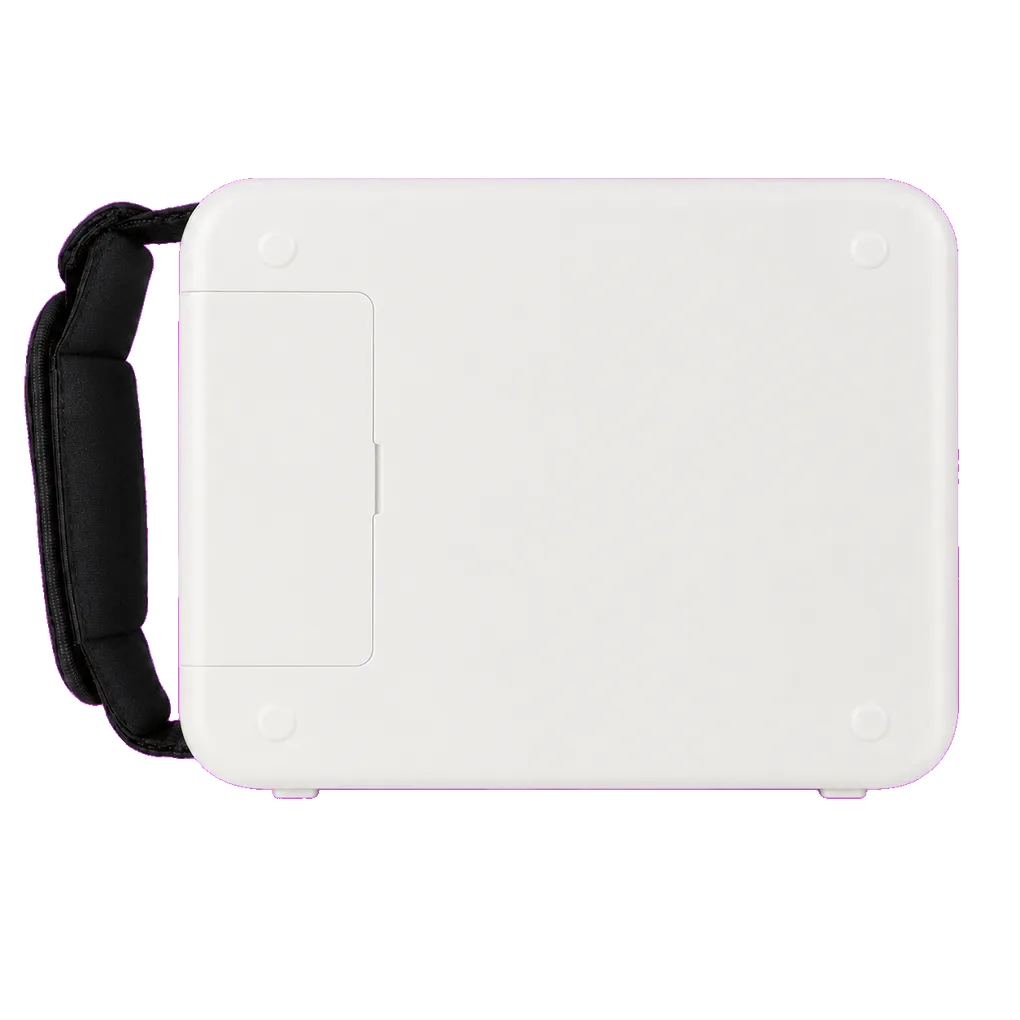
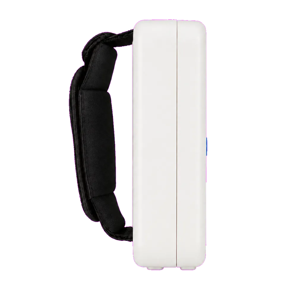
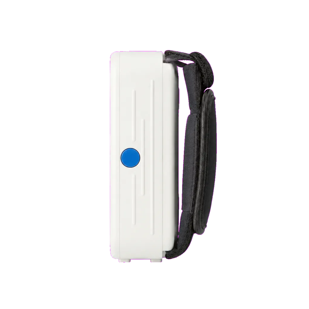
Community and special care teams need equipment one clinician can carry. At 1.7 kg, the unit travels easily, though the modest published battery capacity makes spare pack availability worth settling before a domiciliary round, with off-site radiation protection arrangements agreed with the RPA in advance.
Guidance from the College of General Dentistry and UKHSA Dental X-ray Protection Services remains the reference point UK practices work to, alongside HSE enforcement of IRR17.