
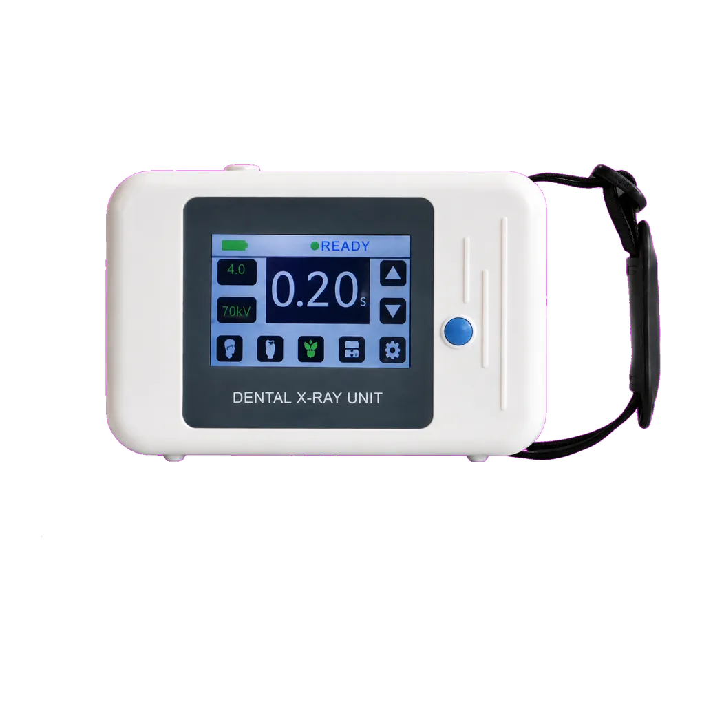
Intraoral radiography carries the diagnostic load in UK general dentistry, and the SJD-141 is configured for that daily volume. It supports bitewing caries detection, periapical assessment of pathology, endodontic working-length and obturation verification, alveolar bone-level review, and root morphology assessment ahead of extraction.
Orthodontic practice is a clear fit. Parallax localisation of unerupted or ectopic canines — two periapical views taken at differing tube angulations to establish palatal or buccal position — is routine, and so is monitoring root resorption through active treatment. Both depend on controlled, repeatable geometry between exposures, which places the emphasis on beam alignment discipline and a documented protocol.
Oral surgery and extraction planning use periapical views to establish root number and curvature, ankylosis, hypercementosis and the relationship of roots to the inferior alveolar canal or maxillary antrum. Those findings change surgical plans and trigger referrals. Where three-dimensional canal relationships are the deciding question, sectional imaging is requested alongside under IR(ME)R justification.
Endodontic practice makes heavy use of periapical views, with a single root canal treatment often needing a diagnostic film, a working-length film, a master cone check and a post-obturation view. Paediatric dentistry benefits from the lower milliamp steps and the 0.1 second timing floor.
Community dental services and special care teams need equipment one clinician can carry. At 1.7 kg, the unit travels easily, though the modest published battery capacity — alongside the standby draw of a backlit panel — makes spare pack availability worth settling before a domiciliary round, with off-site radiation protection arrangements agreed with the RPA in advance.
Guidance from the College of General Dentistry and UKHSA Dental X-ray Protection Services remains the reference point UK practices work to, alongside HSE enforcement of IRR17.