
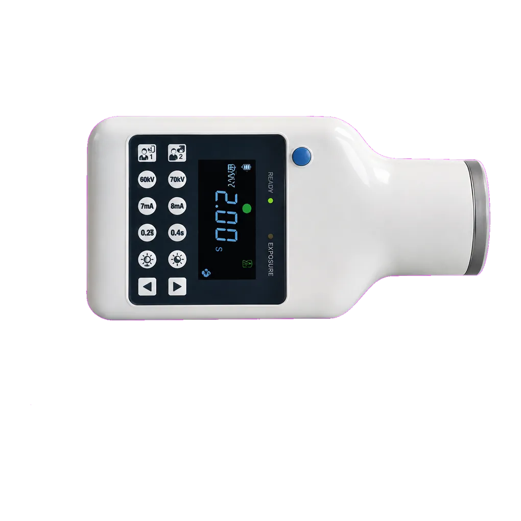
Intraoral radiography carries the diagnostic load in UK general dentistry, and the SJD-221 is configured for that daily volume. It supports bitewing caries detection, periapical assessment of pathology, alveolar bone-level review, and root morphology assessment ahead of extraction.
Endodontic practice is the clearest fit. A single root canal treatment routinely generates a diagnostic periapical, a working-length film, a master cone check, a post-obturation view and a review film at follow-up. Across that sequence the unit's stable output supports comparison from one view to the next, which is what makes serial interpretation meaningful — an apparent change in canal outline or periapical radiolucency should reflect the tooth, not the exposure.
The findings that matter are fine ones. Canal number and curvature, calcification narrowing a chamber with age, pulp stones, internal and external resorption, the proximity of caries to the pulp chamber, and the crown-to-root relationship all shape whether a tooth is treated conservatively, opened, or removed. Radiographic limits should be held in view too — intraoral views are two-dimensional, and a missed canal is a recognised limitation, so small-volume sectional imaging is requested where canal anatomy is the deciding question.
Restorative and prosthetic work uses the same views for crown margin assessment, post space evaluation and crown-to-root ratio before planning. Periodontal practice reads alveolar bone levels on periapicals and bitewings at recall.
Community dental services and special care teams need equipment one clinician can carry. At 1.7 kg the unit travels easily, though the modest published battery capacity is worth pressing on where long endodontic appointments are expected, with off-site radiation protection arrangements agreed with the RPA in advance.
Guidance from the College of General Dentistry and UKHSA Dental X-ray Protection Services remains the reference point UK practices work to, alongside HSE enforcement of IRR17.