
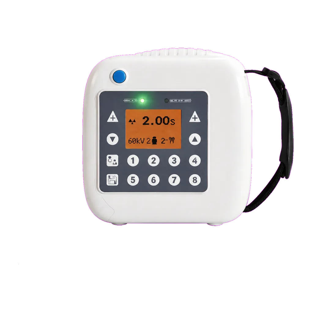
Intraoral radiography carries the diagnostic load in UK general dentistry, and the SJD-331 is configured for that daily volume. It supports bitewing caries detection, periapical assessment of pathology, endodontic length verification, alveolar bone-level review and root morphology assessment ahead of extraction.
Fixed prosthodontics is the application this page is built around. Planning a bridge means assessing each proposed abutment individually — periapical status, root filling quality, bone support, crown-to-root ratio, root form and divergence, existing cores and restorations, and caries at margins. A workup routinely generates several periapical views in one appointment, and the findings decide whether the span is viable, whether the design changes, or whether the case is referred. Post-cementation review films are read against those pre-treatment images.
Upper posterior work brings the maxillary sinus into the picture. Its floor lies close to premolar and molar roots, and the relationship matters before extraction because of oro-antral communication risk. It also complicates interpretation, since the sinus outline can superimpose on apices and either mimic a periapical radiolucency or hide one — a point worth flagging to less experienced operators reading these views.
Restorative and periodontal practice use the same projections for margin assessment, bone-level review at recall and endodontic verification where an abutment needs treatment before it can carry load.
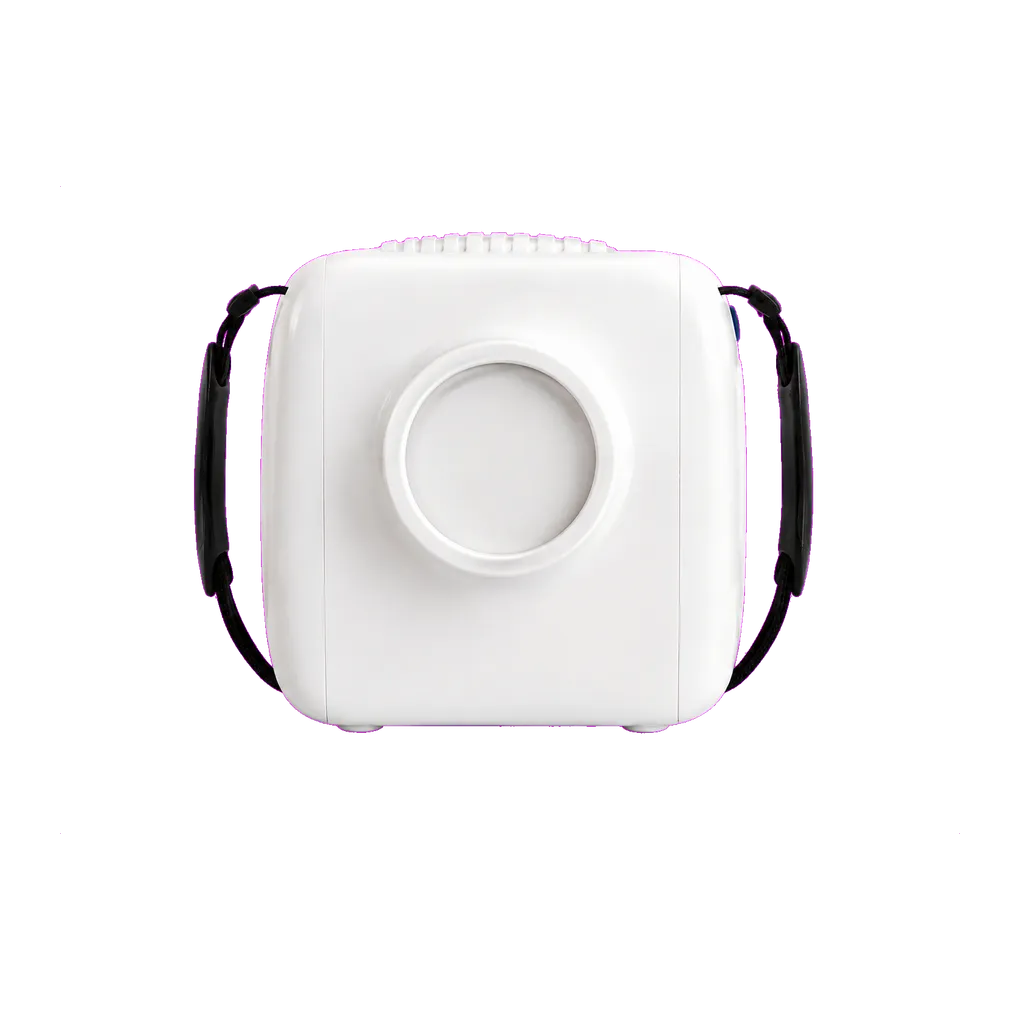
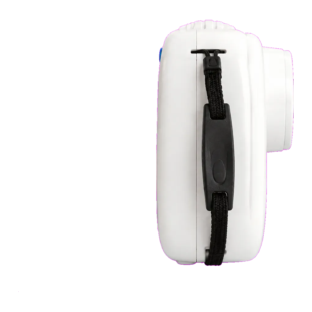
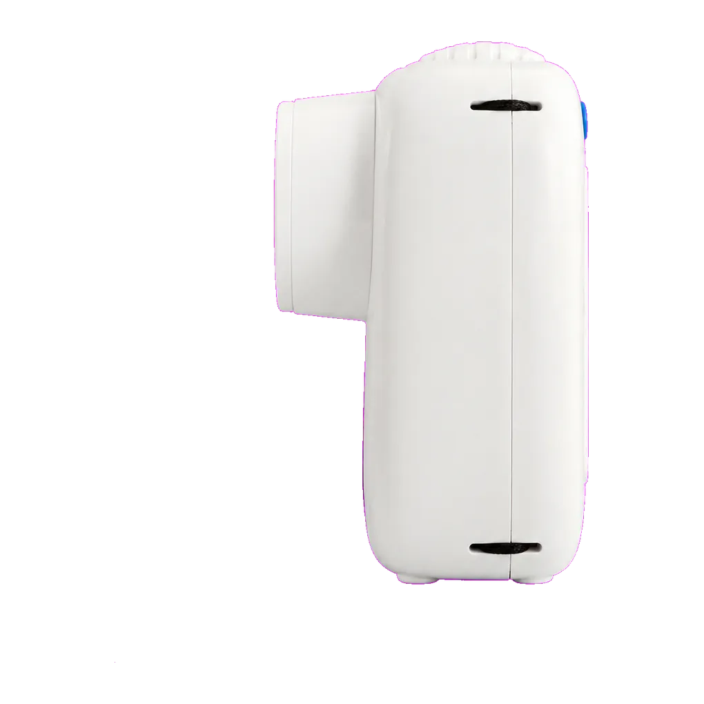
Community dental services and special care teams need equipment one clinician can carry. At 1.7 kg, the unit travels easily, and a 145 × 145 × 140 mm body stows in a lockable drawer between sessions, helping discharge the physical security duty IRR17 places on the employer.
Guidance from the College of General Dentistry and UKHSA Dental X-ray Protection Services remains the reference point that UK practices work to, alongside HSE enforcement of IRR17.