
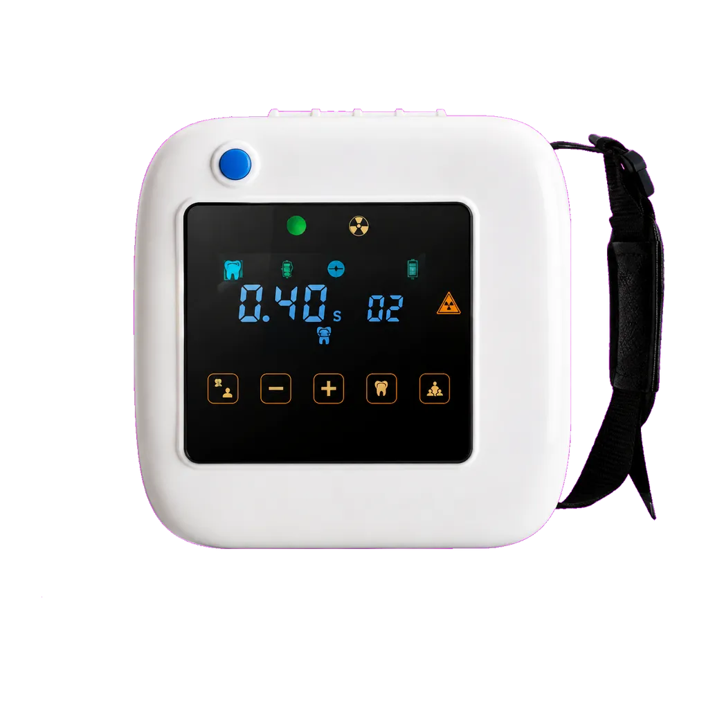
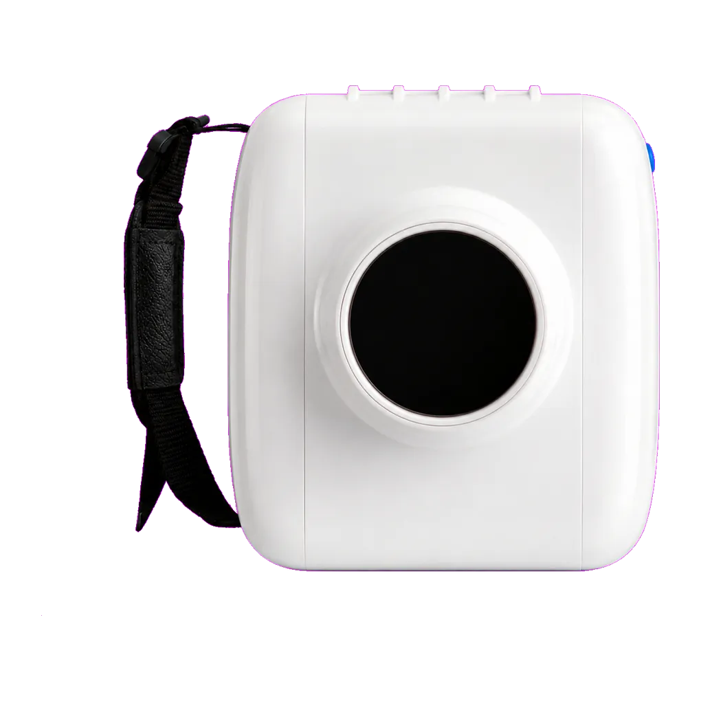
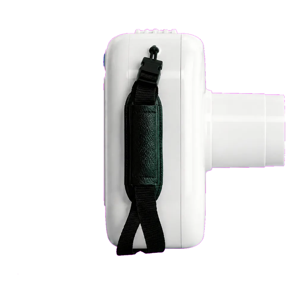
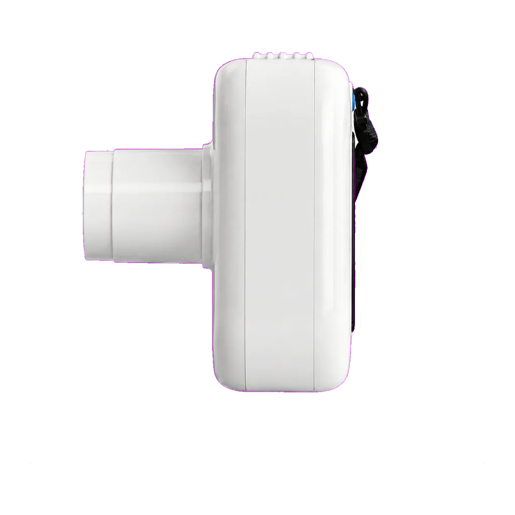
Intraoral radiography carries the diagnostic load in UK general dentistry, and the SJD-341 is configured for that daily volume. It supports bitewing caries detection, periapical assessment of pathology, endodontic length verification, alveolar bone-level review and root morphology assessment ahead of extraction.
Oral surgery gives these projections a defined role. The distal surface of the second molar adjacent to an impacted third molar is the territory that matters most — caries or external resorption there is common and frequently the pathology that justifies removal. NICE guidance on wisdom tooth extraction directs treatment towards teeth with associated pathology rather than routine prophylactic removal, so establishing whether pathology is present is usually the radiographic question, and it is often answered on the neighbouring tooth rather than on the impacted one.
Assessing the impacted tooth itself is a different task. Position, angulation, root morphology and the relationship to the inferior alveolar canal are read from a panoramic radiograph as standard, with small-volume sectional imaging where the canal relationship decides the surgical approach.
Post-operative work is a strong intraoral fit. Socket healing, retained root fragments and any concern localised to a single site read well on a periapical, and a carried unit suits a patient who has just had surgery.
Mandibular views carry an interpretive trap worth naming in training: the mental foramen can appear as a well-defined radiolucency near the lower premolar apices and be mistaken for periapical pathology. A second view at a shifted horizontal angulation separates them, and pulp testing settles it.
Guidance from the College of General Dentistry and UKHSA Dental X-ray Protection Services remains the reference point that UK practices work to, alongside HSE enforcement of IRR17.