
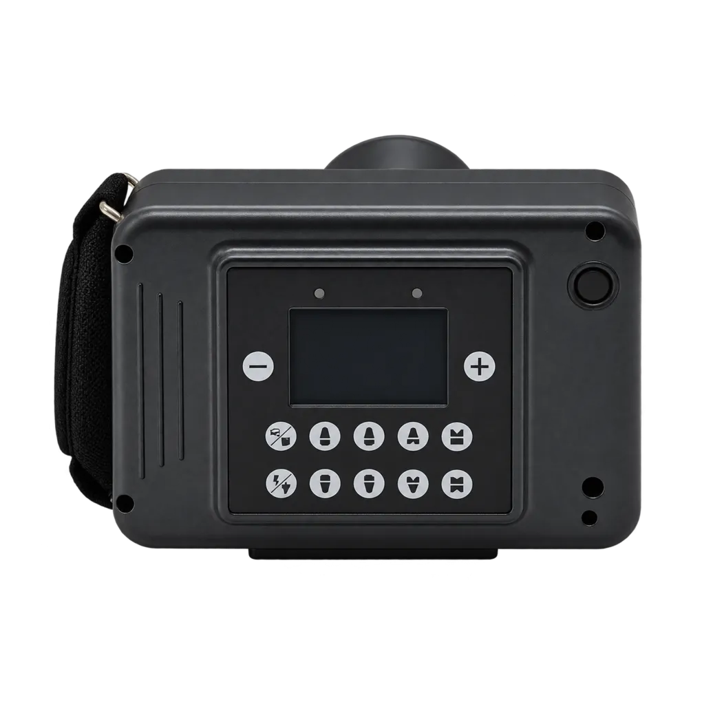
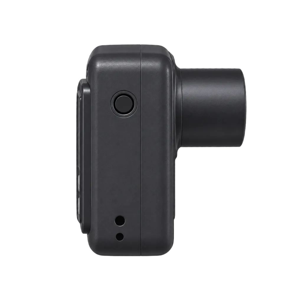
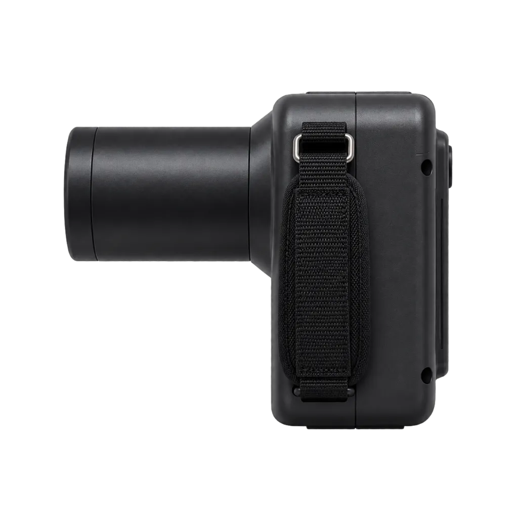
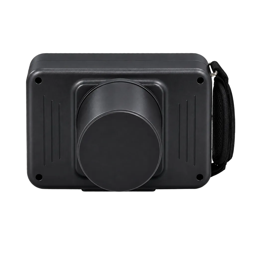
Intraoral radiography carries the diagnostic load in UK general dentistry, and the SJD-C12 is configured for that daily volume. It supports bitewing caries detection, periapical assessment of pathology, endodontic working-length and obturation verification, alveolar bone-level review, and root morphology assessment ahead of extraction.
The geometry of the image shapes what each of those tasks can achieve. Bitewings depend on the beam meeting the line of the contact points squarely, because superimposed proximal surfaces conceal the interproximal areas the view exists to examine. Endodontic working-length films are measured, so the inevitable enlargement between tooth and receptor has to be accounted for through a reference of known length rather than read off the image. Periapical assessment of early bone change depends on edge definition, which is where focal spot size and receptor proximity come together.
Occlusal projections widen the range usefully. They are the standard means of locating an unerupted or ectopic canine in the palate, of demonstrating a salivary calculus in the submandibular duct, and of showing the extent of a cyst or a fracture across an arch. Because occlusal receptors are larger than periapical ones, confirm the X-ray field dimensions with the manufacturer before committing to that work — the field size is not published here.
Paediatric, special care and domiciliary work all benefit from a carried unit reaching the patient, though weight is not published on this data sheet and should be requested before single-person handling is assumed. Off-site radiation protection arrangements need to be agreed with the Radiation Protection Adviser in advance.
Guidance from the College of General Dentistry and UKHSA Dental X-ray Protection Services remains the reference point UK practices work to, alongside HSE enforcement of IRR17.