
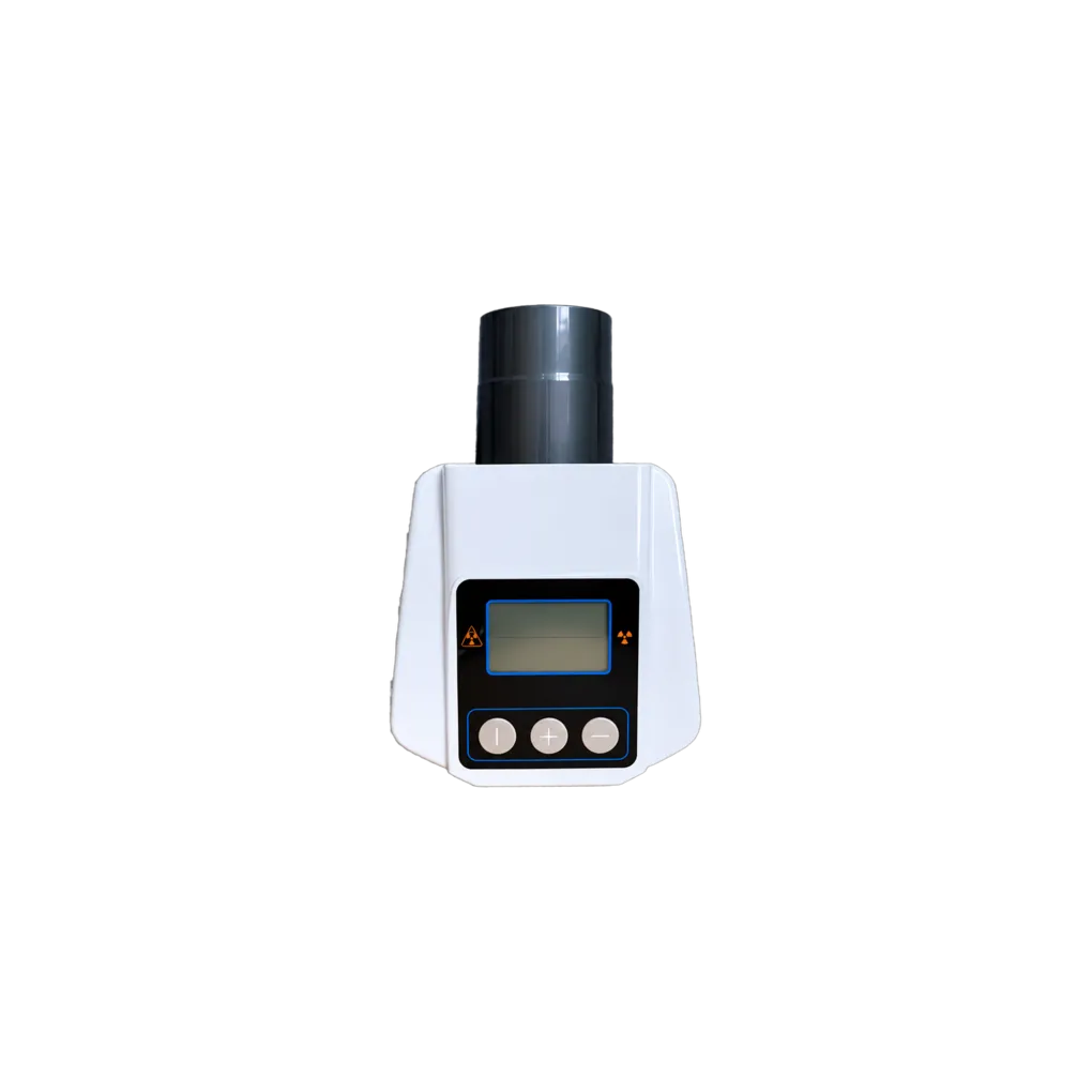
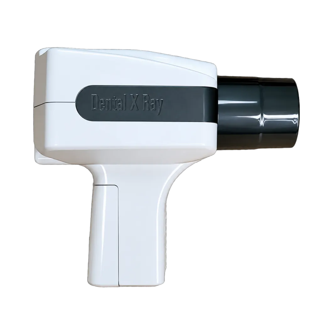
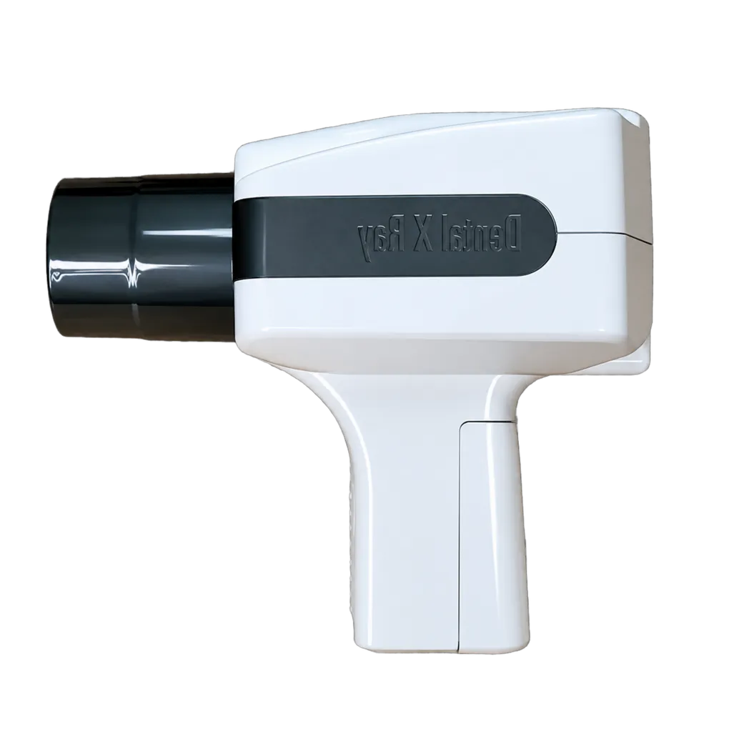
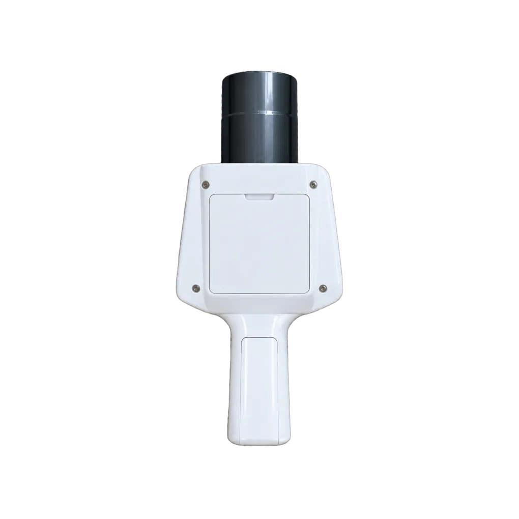
Intraoral radiography carries the diagnostic load in UK general dentistry, and the SJD-PX02 is configured for that daily volume. It supports bitewing caries detection, periapical assessment of pathology, endodontic working-length and obturation verification, alveolar bone-level review, and root morphology assessment ahead of extraction.
What distinguishes this unit clinically is how much of its beam specification is published. Optimisation under IR(ME)R 2017 is the duty to keep doses as low as reasonably practicable while producing images adequate for the diagnostic task, and it runs on figures — total filtration, field size, exposure factors, receptor speed. A data sheet that states total filtration and field dimensions lets a Medical Physics Expert engage with the equipment before it arrives rather than after commissioning.
The 0.01 second exposure step is the practical expression of that. Roughly 130 discrete settings between 0.05 and 1.35 seconds means a protocol can be tuned closely by region, patient build and receptor speed, and optimised downwards in small increments after an image quality audit rather than in coarse jumps.
Collimation is the one specification that deserves scrutiny before purchase. A 50 mm round field sits within the beam diameter limit UK guidance sets at the skin, but rectangular collimation is what the same guidance recommends, because shaping the beam to the receptor removes a substantial share of irradiated tissue. Practices committed to dose optimisation, and services answerable for paediatric exposures in particular, should settle the collimator question at specification.
Community dental services and special care teams need equipment one clinician can carry. At 1.8 kg, the unit travels, with off-site radiation protection arrangements agreed with the RPA in advance.
Guidance from the College of General Dentistry and UKHSA Dental X-ray Protection Services remains the reference point that UK practices work to, alongside HSE enforcement of IRR17.