A D-dimer testing device quantifies a fibrin degradation product that appears in blood as the body dismantles a clot, and that number underpins the safe assessment of suspected deep vein thrombosis and pulmonary embolism. Its clinical power lies in a high negative predictive value: a result below the cut-off, paired with a low Wells or Geneva probability score, lets a clinician exclude venous thromboembolism without sending the patient for imaging. For anyone specifying one, the live questions are the assay chemistry, whether the analyser lives in the coagulation laboratory or at the bedside, which reporting units and cut-off it uses, and the turnaround your rule-out pathway can tolerate.
Treat D-dimer as an exclusion tool rather than a confirmation test. Almost any inflammatory or thrombotic process lifts it, so a raised figure never diagnoses a clot on its own; it simply moves the patient towards imaging.
Why the body produces D-dimer
A stable clot depends on fibrin strands that have been chemically cross-linked into a mesh. When plasmin later dismantles that mesh, it cleaves the cross-linked fibrin and frees D-dimer, a fragment carrying two joined D-domains, which is where the name comes from. Measurable D-dimer therefore tells you that both clot formation and clot breakdown have recently occurred. A level within the reference range makes clinically important thrombosis improbable, which is exactly why the marker earns its place in rule-out algorithms. The catch is specificity: pregnancy, sepsis, recent surgery, cancer, liver disease and advancing age all raise D-dimer, so an elevated value is expected in many unwell patients and cannot stand as proof of a clot.
Inside a D-dimer analyser
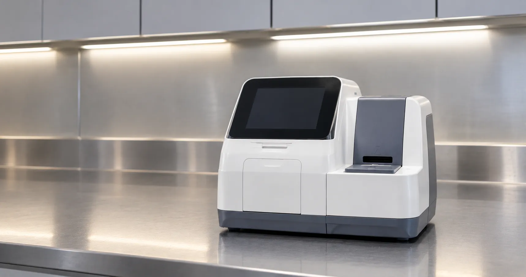
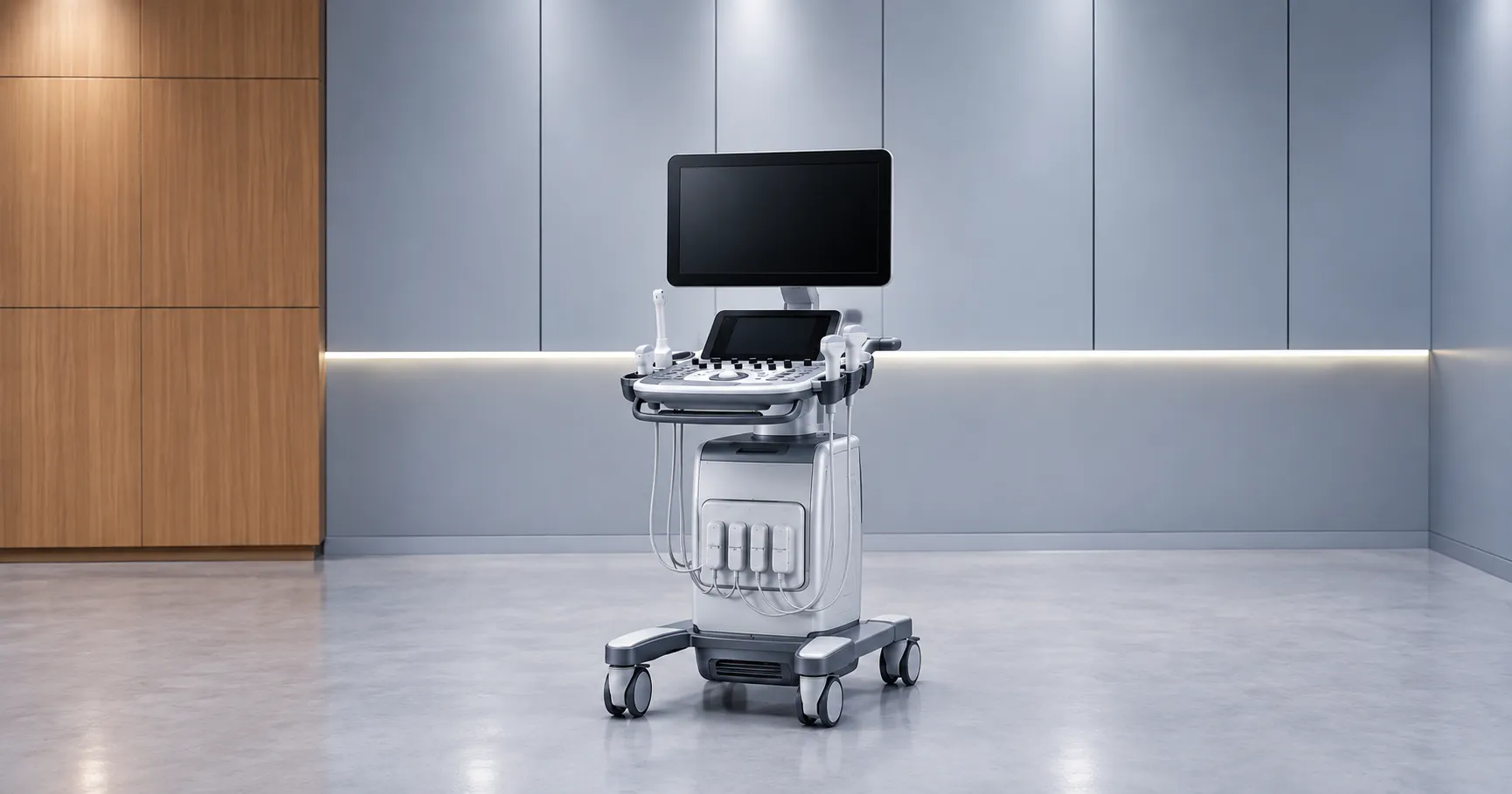
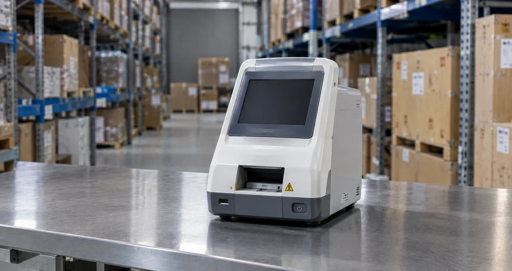
Quantitative platforms rely on antibodies raised against the cross-linked D-dimer fragment. On a coagulation analyser th, the usual method is immuno-turbidimetry: latex microparticles coated with those antibodies agglutinate in proportion to the D-dimer present, and the instrument reads the resulting change in light transmission through the plasma. Near-patient analysers more often run a fluorescence immunoassay on a sealed cartridge, delivering a numeric answer from a drop of whole blood in a few minutes. Legacy qualitative options, such as latex slide agglutination or a whole-blood card giving only positive or negative, lack the graded output the rule-out pathway needs and should not be specified for exclusion work.
Reading the number: FEU, DDU and age-adjusted cut-offs
Nothing trips up a D-dimer procurement more than units. Assays report in one of two conventions, fibrinogen-equivalent units (FEU) or D-dimer units (DDU), and an FEU figure is roughly twice the DDU figure, so the two are not interchangeable, nd a cut-off from one cannot be pasted onto the other. There is no universal calibrator behind these methods, so every threshold is tied to the specific assay's validation data. Many emergency pathways now apply an age-adjusted cut-off above 50 years, typically the patient's age multiplied by ten in nanograms per millilitre FEU, which lifts specificity in older patients without meaningfully harming sensitivity. Before you buy, pin down whether the analyser reports FEU or DDU, whether it can apply age-adjusted logic, and confirm the printed report states the unit so nobody misreads it.
Central lab or bedside: matching the rule-out pathway
Placement is really a question of where the delay sits. A coagulation analyser in the main laboratory processes D-dimer in large batches at a low reagent cost, sitting alongside PT, APTT and fibrinogen, and it suits hospitals whose porters and pneumatic tubes move samples quickly. A cartridge analyser in the emergency department or an urgent treatment centre hands back a quantitative result at the bedside in minutes, which is decisive when transport to the lab is what stretches the rule-out. The bedside route sacrifices batch economy and the keen per-test price of the central bench in exchange for immediacy, and it brings obligations: local governance, trained operators and documented quality control. Verify that the near-patient assay has been validated to the same exclusion standard as your laboratory method, and size capacity for winter surges so a single pending result never stalls a crowded department.
Specifications that decide a D-dimer purchase
Start with the sample type and draw volume, the time to result, and the analytical measuring range, above all its precision around the cut-off, since that is where an exclusion decision is made. Nail down the reporting unit, the validated threshold, and any age-adjusted option. For a bench analyser, weigh throughput and onboard reagent stability; for a cartridge system w, weigh shelf life and storage temperature. Check how the device flags results beyond range, sample interferences such as rheumatoid factor or lipaemia, and control failures, and whether it blocks reporting when controls are overdue. For near-patient siting, judge how easily nursing staff can run it and how cleanly results reach the patient record.
Marking, standards and near-patient governance
Every D-dimer instrument is an in-vitro diagnostic, so it needs UKCA or CE marking with a declaration of conformity, and the manufacturer must appear on the Great Britain registration held by the MHRA. Expect compliance with IEC 61010 for electrical safety and IEC 61326 for electromagnetic compatibility. Laboratory testing runs under ISO 15189, while near-patient placement adds ISO 22870 with named operator competency and clinical sign-off. The way D-dimer slots into venous thromboembolism pathways comes from NICE and local protocol, not from the analyser. Ask to see the conformity file and check the registration entry yourself rather than accepting a brochure line.
Keeping the cut-off trustworthy
When a low D-dimer can send someone home without a scan, control is a patient-safety measure, not a formality. Run at least two levels of quality-control material, one deliberately close to the clinical cut-off, at the maker's stated frequency and after every reagent-lot change; log each value and chase any drift before it reaches a report. Apply assay-specific calibrators when required; most cartridge systems simply scan lot calibration from the pack barcode. Enrol in an external quality assessment scheme, retain the records your ISO 15189 or ISO 22870 accreditation expects, and make sure near-patient sites keep training logs and live connectivity so results and control status flow back to the laboratory for oversight.
What a D-dimer service actually costs to run
The reagent line, not the sticker price, drives the D-dimer business case. A cartridge platform burns one sealed cartridge per test plus collection consumables, whereas a bench analyser draws on bulk reagent, calibrator and control materials whose unit cost drops as batch size grows. Everything that touches blood is single-use and goes to clinical waste, and the housing is wiped with a disinfectant your infection-control team has approved. Model the true cost per reportable result at your real workload, folding in controls and external quality assessment, because a cheap cartridge can conceal short expiry dates or cold-chain storage. Press each supplier on warranty terms, first-year service cover, loan units during breakdowns and the price of predictable failures. The MediGear buyers team can line up configurations side by side, or contact us for stock and support.
Procurement checklist
-
Specify a quantitative immunoassay, never a qualitative latex slide or card, for exclusion work.
-
Confirm FEU or DDU reporting, the validated cut-off and whether age-adjusted logic is available.
-
Scrutinise precision around the cut-off and across the full measuring range.
-
Site the analyser in the lab or at the bedside according to where the delay bites.
-
See the UKCA or CE declaration, IEC 61010 evidence and the MHRA registration entry in writing.
-
Run two control levels, one near the cut-off, backed by external quality assessment.
-
Confirm results and control status reach the laboratory and the patient record.
-
Cost the reportable result across cartridges or bulk reagent, controls included.
Conclusion
A D-dimer analyser earns its keep as a quantitative exclusion aid, and that role stands or falls on precise measurement near the cut-off, an unambiguous reporting unit an, and a threshold validated for the way you use it. Put the instrument wherever your DVT and PE pathway loses time, guard the cut-off with disciplined quality control because a low result can replace a scan, and budget honestly for reagents and quality assessment. Comparing configurations and verified specifications with the MediGear team helps your service land on a D-dimer platform that supports fast, safe thrombosis decisions.
Disclaimer
This article is for informational purposes only. It is published by MediGear (medigear.uk) for general information and procurement guidance, and is not clinical, diagnostic, treatment, technical, engineering, legal or regulatory advice, nor a product endorsement, guarantee or substitute for professional assessment. MediGear does not provide medical consultations. Buyers should consult their clinical, biomedical, estates and regulatory contacts, and the manufacturer's documentation, and independently verify all specifications, certifications, compatibility and suitability before purchase. Specifications, certifications and availability are correct at the time of publication and may change without notice. MediGear is a medical-equipment distributor and does not sell medicines or pharmaceutical products.